Home » Uncategories » Pelvic Anatomy - Pelvic Floor Disorders Anatomy Primal Pictures - Laparoscopic anatomy of the female pelvic region.
Friday, 11 June 2021
Pelvic Anatomy - Pelvic Floor Disorders Anatomy Primal Pictures - Laparoscopic anatomy of the female pelvic region.
Pelvic Anatomy - Pelvic Floor Disorders Anatomy Primal Pictures - Laparoscopic anatomy of the female pelvic region.. Ct body (lymph nodes) ct. Johns hopkins medicine, based in baltimore, maryland This cavity is located within the lesser part of the pelvis, beneath the pelvic brim. The male pelvic floor is a complex structure made up of muscles, ligaments, nerves and fascia. Complete coverage of both conventional and endoscopic surgeries helps you master the full spectrum of surgical procedures.
The pelvic cavity is a body cavity that is bounded by the bones of the pelvis and which primarily contains reproductive organs and the rectum. The male pelvis is different from a female's. The male urethra and the penis Pelvic diaphragm, inferior view (gilroy et al.) atlas of anatomy 2nd ed., fig. Sacrum (the large triangular bone at the base of the spine)
Pelvis Boundaries Springerlink from media.springernature.com {{configctrl2.info.metadescription}} this site uses cookies. A distinction is made between the lesser or true pelvis inferior to the terminal line, and the greater or false pelvis above it. The pelvic cavity is a body cavity that is bounded by the bones of the pelvis and which primarily contains reproductive organs and the rectum. • divided into the true and false pelvis by the iliopectineal line. The male urethra and the penis Sacrum (the large triangular bone at the base of the spine) The pelvic bones are smaller and narrower. The pelvic floor muscles form part of the pelvic floor and play a critical role in sexual function as well as the maintenance of urinary and faecal continence, anatomy of the prostate gland edit
The pelvis is the lower portion of the trunk, located between the abdomen and the lower limbs.
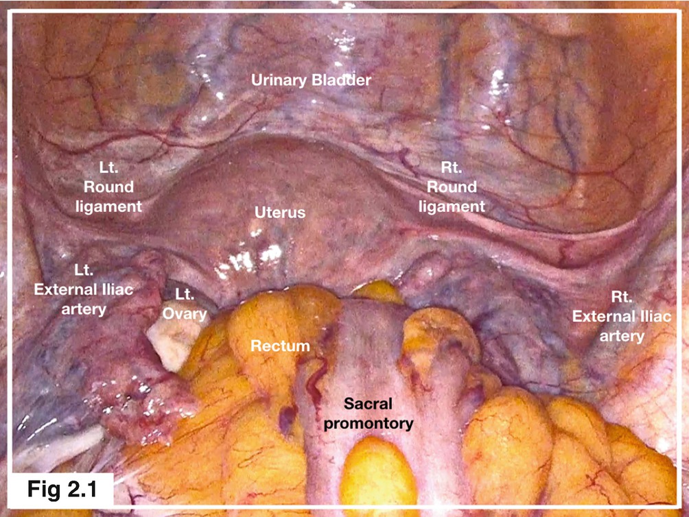
The pelvic region is the area between the trunk — or main body — and the lower extremities, or legs. Ct body (lymph nodes) ct. The pelvis is a musculoskeletal structure that is made up of hip and sacrococcygeal bones, along with several muscular layers. Sacrum (the large triangular bone at the base of the spine) • divided into the true and false pelvis by the iliopectineal line. By continuing to browse this site you are agreeing to our use of cookies. Surgical anatomy of the female pelvis by laparoscopy. Ischial spine ischial tuberosity coccygeus: Laparoscopic anatomy of the female pelvic region. Pelvic floor anatomy, pelvic floor therapy, physical therapist gift, pelvic floor specialist gift, physical therapy office art, pt art. The pelvis's frame is made up of the bones of the pelvis, which connect the axial skeleton to the femurs, and therefore acts in weight bearing of the upper body. The pelvic bones include the: Johns hopkins medicine, based in baltimore, maryland
Covering a compendium of gynecologic operations, including major and minor procedures and approaches, the techniques. The pelvic viscera (bladder, rectum, pelvic genital organs and terminal part of the urethra) reside within the pelvic cavity (or the true pelvis). The pelvis is a basin shaped bony structure formed by the combination of two pelvic bones (hip bones or innominate bones) and the sacrum. The pelvic inlet or superior pelvic aperture, which leads into the lesser pelvis, is bordered by the promontory, the. Laparoscopic anatomy of the female pelvic region.
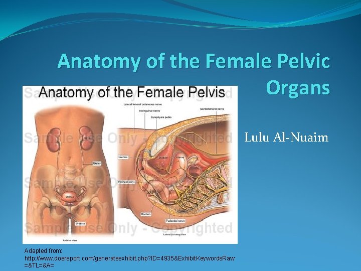
Anatomy Of The Female Pelvic Organs Lulu Alnuaim from slidetodoc.com The pelvis is a basin shaped bony structure formed by the combination of two pelvic bones (hip bones or innominate bones) and the sacrum. The pelvis is a musculoskeletal structure that is made up of hip and sacrococcygeal bones, along with several muscular layers. Each hip bone, in turn, is firmly joined to the axial skeleton via its attachment to the sacrum of the vertebral column. The pelvic viscera (bladder, rectum, pelvic genital organs and terminal part of the urethra) reside within the pelvic cavity (or the true pelvis). Pelvic diaphragm, inferior view (gilroy et al.) atlas of anatomy 2nd ed., fig. The male pelvic floor is a complex structure made up of muscles, ligaments, nerves and fascia. The pelvic bones include the: Surgical anatomy of the female pelvis by laparoscopy.
The pelvis is the lower portion of the trunk, located between the abdomen and the lower limbs.
It is strengthened and supported by several joints and ligaments. It is usually divided into two separate anatomic regions: Each hip bone, in turn, is firmly joined to the axial skeleton via its attachment to the sacrum of the vertebral column. The pelvis is the lower part of the torso. This mri male pelvis axial cross sectional anatomy tool is absolutely free to use. The pelvis is the lower portion of the trunk, located between the abdomen and the lower limbs. {{configctrl2.info.metadescription}} this site uses cookies. Sacrum (the large triangular bone at the base of the spine) This cavity is located within the lesser part of the pelvis, beneath the pelvic brim. Laparoscopic anatomy of the female pelvic region. The pelvic viscera (bladder, rectum, pelvic genital organs and terminal part of the urethra) reside within the pelvic cavity (or the true pelvis). The pelvic floor muscles form part of the pelvic floor and play a critical role in sexual function as well as the maintenance of urinary and faecal continence, anatomy of the prostate gland edit source The testicles and scrotum are also important male structures.
Each hip bone, in turn, is firmly joined to the axial skeleton via its attachment to the sacrum of the vertebral column. Use the mouse scroll wheel to move the images up and down alternatively use the tiny arrows (>>) on both side of the image to move the images.>>) on both side of the image to move the images. The pelvic bones are smaller and narrower. Covering a compendium of gynecologic operations, including major and minor procedures and approaches, the techniques. Laparoscopic anatomy of the female pelvic region.
Anatomy Of The Female Pelvic Organs Lulu Alnuaim from slidetodoc.com Inferior end of sacrum lateral attachment: It provides attachment to some important muscles in the region, and forms a cavity which accommodates several important internal organs. Laparoscopic anatomy of the female pelvic region. The pelvic bones are smaller and narrower. The pelvic girdle (hip girdle) is formed by a single bone, the hip bone or coxal bone (coxal = hip), which serves as the attachment point for each lower limb. The pelvic region is the area between the trunk — or main body — and the lower extremities, or legs. • pelvis begins at the iliac crests and ends at the symphysis pubis. It is further divided into the greater (false) and lesser (true) pelvis.
• pelvis begins at the iliac crests and ends at the symphysis pubis.
The pelvic girdle and pelvic spine. The male urethra and the penis Anatomy the pelvis is a ring of bones located at the lower end of the trunk—between the spine and the legs. The lining of the uterus. The pelvis's frame is made up of the bones of the pelvis, which connect the axial skeleton to the femurs, and therefore acts in weight bearing of the upper body. This area provides support for the intestines and also contains the bladder and reproductive organs. It is further divided into the greater (false) and lesser (true) pelvis. Pelvic floor anatomy, pelvic floor therapy, physical therapist gift, pelvic floor specialist gift, physical therapy office art, pt art. The testicles and scrotum are also important male structures. The pelvic viscera (bladder, rectum, pelvic genital organs and terminal part of the urethra) reside within the pelvic cavity (or the true pelvis). It is strengthened and supported by several joints and ligaments. Ebraheim's educational animated video describes the anatomy of the pelvis, the bony structures, ligaments, muscles, blood supply, and nerves.this video a. Complete coverage of both conventional and endoscopic surgeries helps you master the full spectrum of surgical procedures.
0 Response to "Pelvic Anatomy - Pelvic Floor Disorders Anatomy Primal Pictures - Laparoscopic anatomy of the female pelvic region."

0 Response to "Pelvic Anatomy - Pelvic Floor Disorders Anatomy Primal Pictures - Laparoscopic anatomy of the female pelvic region."
Post a Comment